Littleton Downtown Riverwalk
Enjoying Mt. Washington
Franklin's Lesson For Today
NH Helicopter Rides
FREE Stuff To Do
NH Lupine Photos
Avoiding Romance Scams
Pittsburg NH Profile
Mascoma Lake Profile
Farm To Table Restaurants
Carroll NH (Twin Mtn.) Profile
Colebrook NH Profile
Ben Kilham Profile
Whitefield NH Profile
Clark's Trading Post
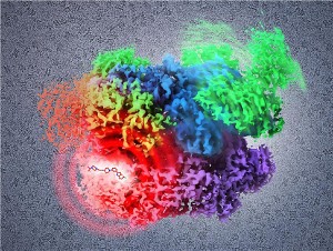
New study using cryo-electron microscopy shows how potential drugs could inhibit cancer.
 The cryo-EM images also helped the researchers establish, at atomic resolution, the sequence of structural changes that normally occur in the protein, p97, an enzyme critical for protein regulation that is thought to be a novel anti-cancer target.
The study appeared online January 28, 2016, in Science. Sriram Subramaniam, Ph.D., of the National Cancer Institute's (NCI) Center for Cancer Research, led the research. NCI is part of the National Institutes of Health.
"Cryo-EM is positioned to become an even more useful tool in structural biology and cancer drug development," said Douglas Lowy, M.D., acting director, NCI. "This latest finding provides a tantalizing possibility for advancing effective drug development."
To determine structures by cryo-EM, protein suspensions are flash-frozen at very low temperatures; nevertheless, the water around the protein molecules stays liquid-like. The suspensions are then bombarded with electrons to capture their images. To produce three-dimensional protein structures using cryo-EM, researchers generate thousands of two-dimensional images of the molecules in different orientations, which are then averaged together. This type of imaging procedure has gained in popularity in structural biology research because it allows for the observation of specimens that have not been stained or fixed in any way, enabling visualization of the specimens under near-native, or natural, conditions.
Earlier structural studies of full-length p97 by a well-established technique known as X-ray crystallography have been limited so far to medium resolution (3.5 Å to 4.7 Å). With cryo-EM, however, researchers were able to image full-length p97 at an overall resolution of 2.3 Å, which is much finer, allowing them to visualize key regions of the protein in atomic detail.
Most significantly, the mode of binding and contact sites of a small molecule inhibitor of p97 activity could be observed directly. Drug development efforts often involve mapping the contacts between small molecules and their binding sites on specific proteins. With this latest finding, the resolutions achieved were significant enough to discern both the shape of the protein chain and some of the hydrogen bonds between the protein and the small molecule inhibitor.
"Our latest research provides new insights into the protein structures and interactions that are critical for the activity of a cancer cell, and this knowledge will hopefully enable the design of clinically useful drugs," said Subramaniam.
Subramaniam and colleagues recently used cryo-EM to understand the functioning of a variety of molecules, including proteins and receptors found in brain cells. The level of detail achieved in their new study however, resolutions of 2.3 Å and 2.4 Å for p97 with and without the bound inhibitor, are second only the 2.2 Å resolution structure reported last year in Science for an enzyme, also by the same group of NCI researchers.
The National Cancer Institute leads the National Cancer Program and the NIH's efforts to dramatically reduce the prevalence of cancer and improve the lives of cancer patients and their families, through research into prevention and cancer biology, the development of new interventions, and the training and mentoring of new researchers. For more information about cancer, please visit the NCI website at www.cancer.gov or call NCI's Cancer Information Service at 1-800-4-CANCER .
Posted 1/31/16
Seussical (9/6-15)
2001 Space Odyssey (9/7)
Muster in the Mountains (9/7-9)
Wingzilla (9/8)
Canzoniere Grecanico Salentino (9/9)
Things [Mom] Taught Me (9/13-23)
Reach the Beach (9/14-15)
WM Storytelling Festival (9/14-16)
Metallak Race (9/15)
Mandeville & Richards (9/15)
Cohase Film Slam (9/16)
Tricycle Grand Prix (9/16)
Rodney Crowell (9/21)
NH Highland Games (9/21-23)
Don Who (9/22)
Harvest Celebration (9/22)
Health & Wellness Fair (9/22)
Jeep Invasion (9/22)
Lakes Region Tri Festival (9/22-23)
Pat Metheny (9/26)
Driving Miss Daisy (9/27 - 10/6)
Neko Case (9/27)
Shot of JD (9/28)
Dixville Half Marathon (9/29)
New Hampshire Marathon (9/29)
Matthew Odell (9/30)
Lincoln Fall Craft Festival (10/6&7)
Sparrow Blue & Crowes Pasture (10/6)
Oktoberfest (10/6-7)
White Mountain Oktoberfest (10/6-7)
Fall Foliage Celebration (10/6-8)
Sandwich Fair (10/6-8)
Paddle the Border (10/7)
Lincoln Fall Craft Festival (10/7-9)
Shadow Play (10/10)
Killer Joe (10/11-21)
Greg Brown (10/12)
Camping & RV Show (10/12-14)
Riverfire & Horrorfest (10/13)
Jay Stollman Band (10/19)
Murder Dinner Train (10/19-20)
Pumpkin Patch Express (10/19-21)
All Things Pumpkin (10/20)
Ethan Setiawan Band (10/20)
Bettye LaVette (10/26)
Murder Dinner Train (10/26-27)
Pumpkin Patch Express (10/26-28)
Berlin Jazz (10/27)
Photo Galleries


Business Directory
Moffett House Museum
Northland Restaurant
Perras Treasures Party Store
Personal Touch Home Health
White Mountain Cottages
more ►


Clark's Trading Post
Cog Railway
Conway Scenic Railroad
Flume
Fort Jefferson Fun Park
Jericho Mountain ATV Park
Kancamagus Highway
Littleton Riverwalk
Lost River Gorge
Mountain Meadow Funplex
Mt. Washington Auto Road
Polar Caves
Santa's Village
Storyland
Vertical Ventures
Whale's Tail Water Park
Woodstock Inn & Brewery
more ►
Community Profiles
Bethlehem
Bretton Woods
Colebrook
Conway
Franconia
Gorham
Hanover
Jackson
Lebanon
Lincoln
Littleton
North Conway
Pittsburg
Plymouth
Twin Mountain
Whitefield
Wolfeboro
more ►
Recreation and Tourism
Farm To Table Restaurants
Hiking To Crash Sites
Little Ski Areas
Local Movie Theaters
Local Performance Theaters
Moose Tours
more ►
Other Resources
NH Cabins & Cottages
NH Census Info
NH Data (OEP)
NH Fishing Reports
NH Foliage Report
NH Hiking Trail Conditions
NH Lottery
NH Movie Guide
NH Road Conditions
NH Ski Reports
NH Snowmobile Trail Reports
NH State Parks
NH Town Officials Directory
NH Weather
Summer Safety Tips
[.pdf]
White Mtn. National Forest
VisitNH.Gov
Copyright 2012-2018 by George C. Jobel , 603-491-4340. All Rights Reserved.


